Showing posts with label Discovery of cells. Show all posts

Saturday, December 7, 2013

Cells,Discovery of cells & Cell theory

Much of the diversity of forms and functions in living organisms results from small atoms being combinedin different ways to form a number of molecules and molecules form macromolecules. Eventually, thesemacromolecules build cells, tissues, organs and finally, an entire organism.

Cells
A cell is the smallest unit of life that can survive and reproduce on its own, given
information in DNA, energy, and raw materials. Some cells live and reproduce
independently. Others do so as part of a multicelled organism.

Discovery of cells
In the middle of the 17th century, one of the pioneers of microscopy, Robert
Hooke (1635–1703), decided to examine a piece of cork tissue with his home-built
microscope. He saw numerous box shaped structures that he thought resembled
row of empty boxes or rooms, so he called them ‘cells’.

Cell theory
Matthias Schleiden and Theodor Schwann, hypothesized that a plant cell is an independent living unit even
when it is part of a plant and both concluded that the tissues of animals as well as plants are composed of
cells and their products. Together, the two scientists recognized that cells have a life of their own even
when they are part of a multicelled body.
Later, physiologist Rudolf Virchow realized that all cells he studied descended from another living cell.
These and many other observations yielded three generalizations that today constitute the cell theory:
1) Every organism is composed of one or more cells
2) Cell is smallest unit having properties of life
3) Continuity of life arises from growth and division of single cells
Thus, Cell theory is that all organisms consist of one or more cells, which are the basic unit of life.

Cell
A cell is the smallest unit that shows the properties of life.
These properties include -
• Can survive on its own or has potential to do so
• Is highly organized for metabolism
• Senses and responds to environment
• Has potential to reproduce

Structure of Cells

Despite their differences, however, all cells share certain organizational and functional features. Every cellhas a plasma membrane. A plasma membrane is selectively permeable, allows only certain materials tocross. All cell membranes, including the plasma membrane, consist mainly of lipids. The plasmamembrane encloses a fluid or jellylike mixture of water, sugars, ions, and proteins called cytoplasm. Some or all of a cell’s metabolism occurs in the cytoplasm, and the cell’s internal components are suspended in it. All cells start out life with DNA, although a few types of cells lose it as they mature.

Cell type
Biologists have categorized cells into two general types: eukaryotic and prokaryotic cells.
The cells of plants, animals, fungi, protozoa, and algae are eukaryotic, and are placed in a category called Eucarya . All eukaryotic cells have their genetic material surrounded by a nuclear membrane forming the cellular nucleus. They also have a large number and variety of complex organelles, each specialized in the metabolic function it performs. In general, they are large in comparison to Prokaryotic cells. These cell types do not have a nuclear membrane; therefore they lack a cellular nucleus. In addition, they display unique chemical and metabolic characteristics but do not have the variety and number of organelles seen in eukaryotes.

Lipid Bilayer
Lipids—mainly phospholipids—make up the bulk of a cell membrane. A phospholipid consists of a phosphate containing head and two fatty acid tails. The polar head is hydrophilic, which means that it interacts with water molecules. The nonpolar tails are hydrophobic, so they do not interact with water molecules, but they do interact with the tails of other phospholipids. Lipid bilayers are the basic structural and functional framework of all cell membranes, gives membrane it's fluidity.


Fluid mosaic

Other molecules, including steroids and proteins, are embedded in or associated with the lipid
bilayer of every cell membrane. Most of these molecules move around the membrane more or less freely. A cell membrane behaves like a two dimensional liquid of mixed composition, so we
describe it as a fluid mosaic. The “mosaic” part of the name comes from a cell membrane’s mixed composition of lipids and proteins. The fluidity occurs because the phospholipids in a cell membrane are not bonded to one another. They stay organized as a bilayer as a result of collective hydrophobic and hydrophilic attractions.

Membrane proteins Separate

Many types of proteins are associated with a cell membrane, and each type adds a specific function to it, different cell membranes can have different characteristics depending on which proteins are associated with them. For example, a plasma membrane has certain proteins that no internal cell membrane has. Many plasma membrane proteins are enzymes. Others are adhesion proteins, which fasten cells together in animal tissues. Recognition proteins function as identity tags for a cell type, individual, or species. Being able to recognize “self” means that foreign cells (harmful ones, in particular) can also be recognized. Receptor proteins bind to a particular substance outside of the cell, such as a hormone or toxin. Binding triggers a change in the cell’s activities that may involve metabolism, movement, division, or even cell death. Receptors for different types of substances occur on different cells, but all are critical for homeostasis. Additional proteins occur on all cell membranes. Transport proteins move specific substances across a membrane, typically by forming a channel through it. These proteins are important because lipid bilayers are impermeable to most substances, including ions and polar molecules. Some transport proteins are open channels through which a substance moves on its own across a membrane.

Movement of Molecules Across the Membrane

Cells must continuously receive nutrients and rid themselves of waste products—one of the characteristics of life. Many of the proteins that are associated with the plasma membrane are involved in moving molecules across the membrane. Some proteins are capable of moving from one side of the plasma membrane to the other and shuttle certain molecules across the membrane. Others extend from one side of the membrane to the other and form channels through which substances can travel. Some of these channels operate like border checkpoints, which open and close when circumstances dictate. Some molecules pass through the membrane passively, whereas others are assisted by metabolic activities within the membrane.
Microscopes
Microscopes allow us to study cells in detail. The ones that use visible light to illuminate objects are called light microscopes. There are two types: Simple and Compound. A more powerful microscope is the Electron microscopes use electrons instead of visible light to illuminate samples. Because electrons travel in wavelengths that are much shorter than those of visible light, electron microscopes can resolve details that are much smaller than you can see with light microscopes. Electron microscopes use magnetic fields to focus beams of electrons onto a sample.
Limitations of Light
• Wavelengths of light are 400-750 nm
• If a structure is less than one-half of a wavelength long, it will not be visible
• Light microscopes can resolve objects down to about 200 nm in size
Electron Microscopy
• Uses streams of accelerated electrons rather than light
• Electrons are focused by magnets rather than glass lenses
• Can resolve structures down to 0.5 nm

Cell size

Almost all cells are too small to see with the naked eye. Why? The answer begins with the processes that keep a cell alive. A living cell must exchange substances with its environment at a rate that keeps pace with its metabolism. These exchanges occur across the plasma membrane, which can handle only so many exchanges at a time. Thus, cell size is limited by a physical relationship called the surface-to-volume ratio. By this ratio, an object’s volume increases with the cube of its diameter, but its surface area increases only with the square. If the cell gets too big, the inward flow of nutrients and the outward flow of wastes across that membrane will not be fast enough to keep the cell alive.

Two Major Cell Types

According to their structure, cells can be of two types:
• Prokaryotes eg. Bacteria
• Eukaryotes eg. Fungi, Plants, Animals
Prokaryotic
Prokaryotic cells are so called because they have no nucleus (‘prokaryote’ comes from the Greek,
meaning ‘before the nucleus’). They also have no organelles (internal structures), so there is little
compartmentalization of function within them. From the mid-20th century, when the electron microscope
was developed, it became possible to study the internal detail of cells.
• The cell wall surrounds the cell. It protects the cell from bursting and is composed of peptidoglycan,
which is a mixture of carbohydrate and amino acids.
• The plasma membrane controls the movement of materials into and out of the cell. Some substances
are pumped in and out using active transport.
• Cytoplasm inside the membrane
contains all the enzymes for the
chemical reactions of the cell. It also
contains the genetic material.
• The chromosome is found in a
region of the cytoplasm called the
nucleoid. The DNA is not contained
in a nuclear envelope and also it is
‘naked’ – that is, not associated with
any proteins. Bacteria also contain
additional small circles of DNA called plasmids. Plasmids replicate
independently and may be passed
from one cell to another.
• Ribosomes are found in all prokaryotic cells, where they synthesize proteins. They can be seen in very
large numbers in cells that are actively producing protein.
• A fagellum is present in some prokaryotic cells. A flagellum, which projects from the cell wall, enables a cell to move.
• Some bacteria have pili (singular pilus). These structures, found on the cell wall, can connect to other bacterial cells, drawing them together so that genetic material can be exchanged between them.
Prokaryotic cells are usually much smaller in volume than more complex cells because they have no
nucleus. Their means of division is also simple. As they grow, their DNA replicates and separates into two different areas of the cytoplasm, which then divides into two. This is called binary fission. It differs slightly from mitosis (a type of cell division) in eukaryotic cells.

Eukaryotes
The cells of plants, animals, fungi, protozoa, and algae are eukaryotic, and are placed in a category called Eucarya . All eukaryotic cells have their genetic material surrounded by a nuclear membrane forming the cellular nucleus. They also have a large number and variety of complex organelles, each specialized in the metabolic function it performs. In general, they are large in comparison to prokaryotic cells.

Animal cells
• Plasma membrane
• Nucleus
• Ribosomes
• Endoplasmic reticulum
• Golgi body
• Vesicles
• Mitochondria
• Cytoskeleton

Plant cells
• Plasma membrane
• Nucleus
• Ribosomes
• Endoplasmic reticulum
• Golgi body
• Vesicles

Functions & Components of Nucleus

Functions of Nucleus
• Keeps the DNA molecules of eukaryotic cells separated from metabolic machinery of cytoplasm
• Makes it easier to organize DNA and to copy it before parent cells divide into daughter cells

Components of Nucleus
– Nuclear envelope
– Nucleoplasm
– Nucleolus
– Chromosome
– Chromatin

Nucleus:
The nucleus is the defining organelle of eukaryotic cells. The nucleus is separated from the cytoplasm by a double membrane (two phospholipid bilayers); known as the nuclear envelope. The nuclear envelope controls the passage of molecules between the nucleus and cytoplasm. The nucleus contains the DNA, the stored genetic instructions of each cell. In addition, important reactions for interpreting the genetic instructions occur in the nucleus.
  • In the nucleus, DNA is organized into discrete units called chromosomes
  • Each chromosome is composed of a single DNA molecule associated with proteins
  • The nucleolus is located within the nucleus and is the site of ribosomal RNA (rRNA) synthesis
Nucleolus
  •  Dense mass of material in nucleus
  •  May be one or more
  •  Cluster of DNA and proteins
  •  Materials (mostly rRNA) from which ribosomal subunits are built
  • Subunits must pass through nuclear pores to reach cytoplasm

Chromatin

Chromatin is composed of long molecules of DNA, along with proteins. Most of the time, the chromatin is arranged as a long, tangled mass of threads in the nucleus. However, during cell division, the chromatin becomes tightly coiled into short, dense structures called chromosomes (chromo=color; some=body). Chromatin and chromosomes are really the same molecules, but they differ in structural arrangement. In addition to chromosomes, the nucleus may also contain one, two, or several nucleoli. A nucleolus is the site of ribosome manufacture. Specific parts of the DNA become organized within the nucleus to produce ribosomes. A nucleolus is composed of this DNA, specific granules, and partially completed ribosomes.
  1.  The DNA and proteins of chromosomes are together called chromatin
  2. Chromatin condenses to form discrete chromosomes as a cell prepares to divide
  3. Chromosome is one DNA molecule and its associated proteins
  4. Appearance changes as cell divides

Mitochondria

The mitochondrion (plural, mitochondria) is a type of organelle that specializes in making ATP (molecule used by cells as main energy source). They have various enzymes to catalyze cellular respiration. Bacteria have no mitochondria; they make ATP in their cell walls and cytoplasm. Cells that have a very high demand for energy tend to have many mitochondria e.g. liver needs more because needs more energy. Mitochondria, like most organelles, can move within the cell and they grow and divide independently. Each has two membranes, one highly folded inside the other. Double-membrane system: Smooth outer membrane (lipid bilayer) faces cytoplasm and permeable to small solutes; blocks macromolecules where as Inner Membrane (cristae) folds back on itself to enlarge surface area for chemical reactions to take place. Membranes form two distinct compartments. ATP-making machinery is embedded in the inner mitochondrial membrane.

  • Mitochondria and chloroplasts have similarities with bacteria,
  • Enveloped by a double membrane
  • Contain free ribosomes and circular DNA molecules
  • Grow and reproduce somewhat independently in cells
They may have evolved from ancient bacteria that were engulfed but not digested. Mitochondria and chloroplasts developed because as a prokaryote it gained protection by living inside the eukaryote and in turn produced energy for the eukaryote (symbiotic relationship).

Chloroplasts: Capture of Light Energy

Plastids are a category of membrane-enclosed organelles that function in photosynthesis or storage in
plant and algal cells. Plastids called chloroplasts are organelles specialized for photosynthesis. Chloroplastscontain the green pigment chlorophyll, as well as enzymes and other molecules that function in photosynthesis. Chloroplasts are found in leaves and other green organs of plants and in algae.

Chloroplast structure includes 

Stroma: Each has two outer membranes enclosing a semifluid interior, the stroma, that contains
enzymes and the chloroplast’s own DNA.

Thylakoids: Inside the stroma, a third, highly folded membrane forms a single, continuous compartment. The folded membrane resembles stacks of flattened disks. The stacks are called grana (singular, granum). Photosynthesis takes place at this membrane, which is called the thylakoid membrane. The abundance of chlorophylls in thylakoids is the reason most plants are green. By the process of photosynthesis, chlorophylls and other molecules in the thylakoid membrane harness the
energy in sunlight to drive the synthesis of ATP. The ATP is then used inside the stroma to build carbohydrates from carbon dioxide and water.

Ribosomes: Protein Factories

Ribosomes are nonmembranous organelles responsible for the synthesis of proteins from amino acids. They are composed of RNA and protein. Each ribosome is composed of two subunits—a large one and a small one. As mentioned before, they are constructed in the Nucleolus. Ribosomes carry out protein synthesis in two locations

Bound ribosomes: Many ribosomes are attached to the endoplasmic reticulum. Because ER that has attached ribosomes appears rough when viewed through an electron microscope it is called rough ER. Areas of rough ER are active sites of protein production.


Free ribosomes: Many ribosomes are also found floating freely in the cytoplasm wherever proteins are being assembled. Cells that are actively producing protein (e.g., liver cells) have great numbers of free and attached ribosomes. Ribosomes are not surrounded by membrane (found in prokaryotic cells too)

Cytomembrane System

The cytomembrane system is a series of interacting organelles between the nucleus and the plasma
membrane. Its main function is to make lipids, enzymes, and proteins for secretion, or for insertion into cell membranes. It also destroys toxins, recycles wastes, and has other specialized functions. The
system’s components vary among different types of cells, but here we present the most common ones:
Components of Cytomembrane System
– Endoplasmic reticulum
– Golgi bodies
Vesicles

Endoplasmic Reticulum

Part of the cytomembrane system is an extension of the nuclear envelope called endoplasmic reticulum, or ER. ER forms a continuous compartment that folds into flattened sacs and tubes. The space inside the compartment is the site where many new polypeptide chains are modified. Two kinds of ER, rough and smooth, are named for their appearance in electron micrographs. Thousands of ribosomes are attached to the outer surface of rough ER.
Rough ER
  •  Arranged into flattened sacs
  •  Ribosomes on surface give it a rough appearance
  •  Some polypeptide chains enter rough ER and are modified
  •  Cells that specialize in secreting proteins have lots of rough ER
Smooth ER
  •  A series of interconnected tubules
  •  No ribosomes on surface
  •  Lipids assembled inside tubules
  •  Smooth ER of liver inactivates wastes, drugs
  •  Sarcoplasmic reticulum of muscle is a specialized form that stores calcium

Functions of Smooth & Rough ER

• The smooth ER
1. Synthesizes lipids
2. Metabolizes carbohydrates
3. Detoxifies drugs and poisons
4. Stores calcium ions

• The rough ER
1. Has bound ribosomes
2. Distributes transport vesicles,
proteins surrounded by membranes
3. Is a membrane factory for the cell

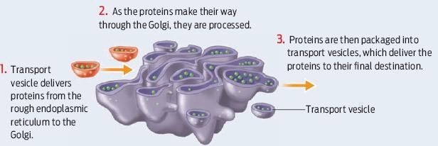
Golgi Bodies

Golgi : The Golgi is a series of flattened membrane compartments, whose purpose is to process and
package proteins produced in-the rough endoplasmic reticulum. The processed molecules are packaged into membrane vesicles, then targeted and transported to-their final destinations.
Functions of the Golgi apparatus
• Modifies products of the ER
• Manufactures certain macromolecules
• Sorts and packages materials into transport vesicles

Vesicles

Small, membrane-enclosed, saclike vesicles form in great numbers, in a variety of types, either on their own or by budding. There are many types but two main are:
 Lysosomes: Digestion & recycling centers


Lysosomes that bud from Golgi bodies take part in intracellular digestion. They contain powerful enzymes that can break down carbohydrates, proteins, nucleic acids, and lipids. Vesicles inside white blood cells or amoebas deliver ingested bacteria, cell parts, and other debris to lysosomes for destruction. The enzymes work best in the acidic environment inside the lysosome. Lysosomes break down worn out cell parts or molecules so they can be used to build new cellular structures. Some types of cell can engulf another cell by phagocytosis; this forms a food vacuole. A lysosome fuses with the food vacuole and digests the molecules Lysosomes also use enzymes to recycle the cell’s own organelles and macromolecules, a process called autophagy

Peroxisomes: In plants and animals, vesicles called peroxisomes form and divide on their own, so they are not part of the endomembrane system. Peroxisomes contain enzymes that digest fatty acids and amino acids. They also break down hydrogen peroxide, a toxic byproduct of fatty acid metabolism. Peroxisome enzymes convert hydrogen peroxide to water and oxygen, or use it in reactions that break down alcohol and other toxins.

Friday, December 6, 2013

The cytoskeleton is a network of fibers that organizes structures and activities in the cell

Between the nucleus and plasma membrane of all eukaryotic cells is a system of interconnected protein filaments collectively called the cytoskeleton. The cytoskeleton is a network of fibers extending throughout the cytoplasm. Elements of the cytoskeleton reinforce, organize, and move cell structures, anchoring many organelles.

Microtubules

Microtubules are long, hollow cylinders that consist of subunits of the protein tubulin. They form a dynamic scaffolding for many cellular processes, rapidly assembling when they are needed and then disassembling when they are not. For example, before a eukaryotic cell divides, microtubules assemble, separate the cell’s duplicated chromosomes, then disassemble. As another example, microtubules that form in the growing end of a young nerve cell support and guide its lengthening in a particular direction.


Microfilaments

Microfilaments are fibers that consist primarily of subunits of the globular protein actin. They strengthen or change the shape of eukaryotic cells. Crosslinked, bundled, or gel-like arrays of them make up the cell cortex, which is a reinforcing mesh under the plasma membrane. Actin microfilaments that form at the edge of a cell drag or extend it in a certain direction. Myosin and Actin microfilaments interact to bring about contraction of muscle cells.

Intermediate filaments

Intermediate filaments that support cells and tissues are the most stable elements of the cytoskeleton. These filaments form a framework that lends structure and resilience to cells and tissues. Some kinds underlie and reinforce membranes. The nuclear envelope, for example, is supported by an inner layer of intermediate filaments called lamins. Other kinds connect to structures that lock cell membranes together in tissues.


Microtubules control the beating of cilia and flagella, locomotor appendages of some cells. Cilia and flagella differ in their beating patterns Cilia – Cilia (singular, cilium) are short, hairlike structures that project from the surface of some cells. Cilia are usually more profuse than flagella. The coordinated waving of many cilia propels cells through fluid, and stirs fluid around stationary cells. Flagella – Eukaryotic flagella are structures that whip back and forth to propel cells such as sperm through fluid. They have a different internal structure and type of motion than flagella of bacteria.

Plant Cells

Plants are eukaryotes and have the typical eukaryotic cell organization, consisting of nucleus and cytoplasm. The cytoplasm is enclosed by a plasma membrane and contains numerous membrane-enclosed organelles, including plastids, mitochondria, microbodies, oleosomes, and a large central vacuole. Chloroplasts and mitochondria are semiautonomous organelles that contain their own DNA.

 The main Characteristics are given below: 
Cell wall – rigid, support & protect plant, Cellulose fiber embedded

Vacuoles – fluid-filled; store enzymes & metabolic wastes

Plastids – contain DNA surrounded by 2 membranes. Store starch/fats

Absorb visible light – pigments

Chloroplast – site where photosynthesis takes place

Thylakoids – membranous sacs contains chlorophyll


Plants have & we don’t:

·         Cell wall

·         Vacuoles

·         Plastids (where photosynthesis takes place)

Cell Walls of Plants


·         The cell wall is an extracellularstructure that distinguishes plant cells from animal cells, made of cellulose fibers embedded in other polysaccharides and protein.

·         Prokaryotes, fungi, and some protists also have cell walls.

·         The cell wall protects the plant cell, maintains its shape, and prevents excessive uptake of water.

The Extracellular Matrix (ECM) of Animal Cells


Most cells of multicelled organisms are surrounded and organized by a nonliving, complex mixture of fibrous proteins and polysaccharides called extracellular matrix, or ECM. Secreted by the cells it surrounds, ECM supports and anchors cells, separates tissues, and functions in cell signaling. 
Different types of cells secrete different kinds of ECM. The cell wall around the plasma membrane of plant cells is a type of ECM that is structurally different from the cell wall of bacteria and archaeans. Both types of wall protect, support, and impart shape to a cell. Both are also porous: Water and solutes easily cross it on the way to and from the plasma membrane. Cells could not live without exchanging these substances with their environment. Plant and animals secrete substances such as collagen, proteoglycans, lignin and fibronectin with their ECM. Cells send and receive ions, molecules, or signals through some junctions. ECM proteins bind to receptor proteins in the plasma membrane called integrins.

Membrane structure results in selective permeability

A cell must exchange materials with its surroundings, a process controlled by the plasma membrane. Plasma membranes are selectively permeable, regulating the cell’s molecular traffic. Hydrophobic (nonpolar) molecules, such as hydrocarbons, can dissolve in the lipid bilayer and pass. Through the membrane rapidly and polar molecules, such as sugars, do not cross the membrane easily.

Transport proteins

Transport proteins allow passage of hydrophilic substances across the membrane Some transport proteins, called channel proteins, have a hydrophilic channel that certain molecules or ions can use as a tunnel. Channel proteins called aquaporins facilitate the passage of water. Other transport proteins, called carrier proteins, bind to molecules and change shape to shuttle them across the membrane. A transport protein is specific for the substance it moves.

Passive transport is diffusion of a substance across a membrane

Diffusion is the tendency for molecules to spread out evenly into the available space.
Although each molecule moves randomly, diffusion of a population of molecules may be directional.
At dynamic equilibrium, as many molecules cross the membrane in one direction as in the other.


Substances diffuse down their :

concentration gradient, the region along which the density of a chemical substance increases or decreases. No work must be done to move substances down the concentration gradient. The diffusion of a substance across a biological membrane is passive transport because no energy is expended by the cell to make it happen. Substances down the concentration gradient. The diffusion of a substance across a biological membrane is passive transport because no energy is expended by the cell to make it happen.

Osmosis and H2O balance of Cells


Osmosis is the diffusion of water across a selectively permeable membrane. Water diffuses across a membrane from the region of lower solute concentration to the region of higher solute concentration until the solute concentration is equal on both sides.


H2O balance of Cells


Tonicity is the ability of a surrounding solution to cause a cell to gain or lose water.
Isotonic solution: Solute concentration is the same as that inside the cell; no net water movement across the plasma membrane.
Hypertonic solution: Solute concentration is greater than that inside the cell; cell loses water.
Hypotonic solution: Solute concentration is less than that inside the cell; cell gains water.
Hypertonic or hypotonic environments create osmotic problems for organisms.